X Ray Machine Explained
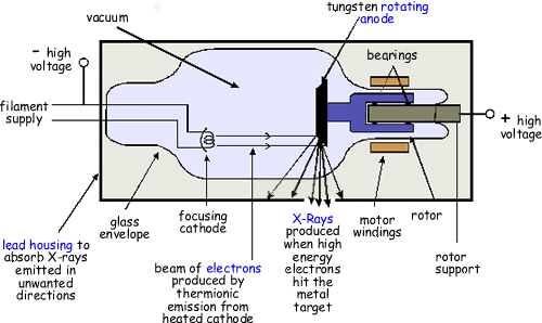
A circuit for heating the filament. The physics of x-ray production will be discussed later in section 34.
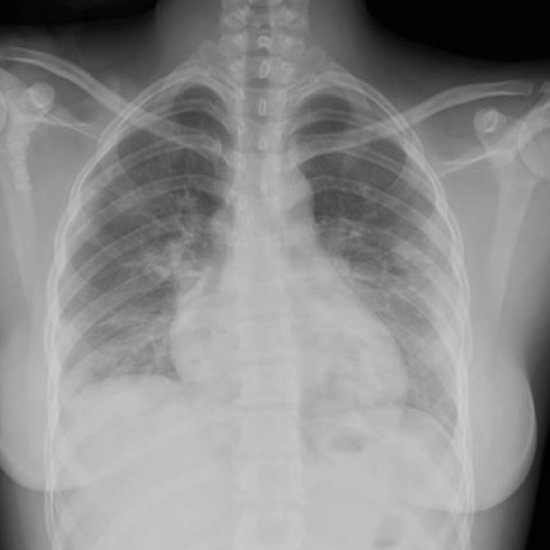
Plain Film X Ray Principles Interpretation Teachmeanatomy
American physicist William David Coolidge 18731975 develops the practical X-ray-making machine.
X ray machine explained. The x-ray machine takes the image from beneath the chin for a view of the lower teeth and jaw or from above near the nose for the upper teeth and jaw. A timing device to control the length of exposure. No external radioactive material is involved.
Introduction to the parts of x ray machine 1. Different X-ray beam spectra are applied to different body parts. The heart of an X-ray machine is an electrode pair -- a cathode and an anode -- that sits inside a glass vacuum tube.
Their equipment features the QS-500 tubestand the Verti-Q vertical wall stand and a variety of radiographic tables. Boost the voltage to the necessary range of x-ray production. The x-rays are produced by the sudden deflection or acceleration of the electron caused by the attractive force of the tungsten nucleus.
X-ray machine circuits comprise three main components. For those needing flexibility and cost-saving pricing Quantum RAD-X meets the need for outstanding hospital x ray equipment. X-ray powder diffraction XRD is a rapid analytical technique primarily used for phase identification of a crystalline material and can provide information on unit cell dimensions.
Permit the radiographer to adjust technical factors. X-rays are produced within the X-ray machine also known as an X-ray tube. Known as a Coolidge tube its a long glass jar with an electron beam and a metal target inside.
The machine passes current through the filament heating it up. How does an X-ray work. An X-ray is a diagnostic test that uses radiation waves called x-rays to take pictures of your body tissues.
A circuit for applying a large potential difference high voltage between cathode and anode to accelerate electrons. Incorporate appropriate circuitry to increase x-ray production efficiency. Huzaifa Atique Sir Syed University of Engineering and Technology 2.
Radiographers can change the current and voltage settings on the X-ray machine in order to manipulate the properties of the X-ray beam produced. Fundamental Principles of X-ray Powder Diffraction XRD. As an X-ray beam passes through your body the body tissues and bones absorb andor block the beam in varying amounts depending on its density.
Supplies power to the x-ray tube so that x-rays are produced. Tube Head or Protective Housing. About Press Copyright Contact us Creators Advertise Developers Terms Privacy Policy Safety How YouTube works Test new features Press Copyright Contact us Creators.
Occlusal x-rays are most commonly used by pediatric dentists to check on the growth and formation of the teeth and jaw bone. X-Ray tubes receive the high frequency line power from the generator and actually create the radiation waves by colliding electrons into a tungsten plate. The basic tube that works well for most general practitioners with a fairly low study volume is a 10.
The analyzed material is finely ground homogenized and average bulk composition is determined. The cathode is a heated filament like you might find in an older fluorescent lamp. The x-ray beam emerges through a thin glass window in the tube envelope.
Vacuum Protection housing Projectile electron 4. Modifies incoming current to produce x-rays. The heat sputters electrons off of the filament surface.
When the beam is fired at the target X rays are produced.

How Do X Rays Work Independent Imaging

Hypocycloidal X Ray Machine Google Search Math X Ray Radiology

X Ray Vision An Accidental Discovery That Revolutionized Medicine Science In Depth Reporting On Science And Technology Dw 06 11 2015
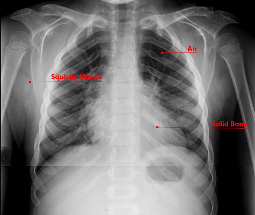
What Is An X Ray For Kids Radiology And Medical Imaging

X Ray Artifacts Radiology Reference Article Radiopaedia Org
Plain Radiograph X Ray Insideradiology

The Facts About Nuclear Medicine Nuclear Medicine Nuclear Medicine Imaging Medicine

Imaging The Coronavirus Disease Covid 19
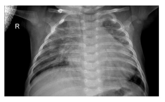
Sensors Free Full Text Detecting Pneumonia Using Convolutions And Dynamic Capsule Routing For Chest X Ray Images Html
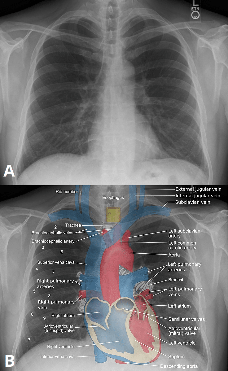
Plain Film X Ray Principles Interpretation Teachmeanatomy

The Working And Benefits Of Dual Source Ct Scan Explained Siemens Scanners Scan Design
X Rays Undergraduate Diagnostic Imaging Fundamentals

X Rays Definition Of X Rays By Medical Dictionary Radiology Humor Radiology Student Radiology Schools

X Ray Imaging Physics For Nuclear Medicine Technologists Part 1 Basic Principles Of X Ray Production Journal Of Nuclear Medicine Technology

Detecting Covid 19 In X Ray Images With Keras Tensorflow And Deep Learning Pyimagesearch

Chest X Ray Interpretation Explained Clearly How To Read A Chest Xray Youtube
X Rays Undergraduate Diagnostic Imaging Fundamentals

You Ve Got A Friend In Me Indeed X Ray Buzz Lightyear Action Figure Buzz Lightyear

Post a Comment for "X Ray Machine Explained"